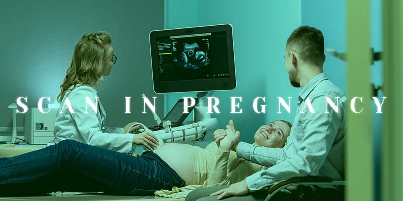
ULTRASOUND SCANS:
Ultrasound scans in pregnancy are commonly used scans in pregnancy The pregnancy span for women is around 40 weeks starting from the first day of the mother’s last menstrual period. The scans are usually done by Fetal medicine specialists, Radiologists, Obstetricians, and Sonographers. In ultrasound scans, high-frequency sound waves are used to create an image of the baby in the womb. USG is used to track the baby’s growth. Every pregnant woman must know about USG and its types in detail to know their baby’s growth better. Let’s see the USG in detail in this blog.
It is an important noninvasive magic wand in the hands of the obstetrician.USG focuses high-energy sound waves on the uterus and gets an image signal as grey and white(2D). It helps in assessing the proper growth and development of the baby, anomalies, liquor, and location of the placenta. Let’s get deep into the three most important scans done during pregnancy.
DATING SCAN:
Primarily,self-confirm your pregnancy by card test. When the result is positive, go ahead and consult your obstetrician for further confirmation through scanning. The dating scan is also known as the Viability scan. It is the first scan done at 6 to 8 weeks of pregnancy. A viability scan is done for the following reasons
*To confirm pregnancy
*To find the number of fetuses
*To find the type of twinning in case of multiple pregnancies (Chorionicity).
*To find the cardiac activity and gestational age
The Estimated Date of Delivery(EDD) can be fixed based on this scan.
NT SCAN:
It is an important scan done at the 11 to 13 weeks 6 days of pregnancy . This scan helps to detect major anomalies in the brain and spinal cord of the baby. It provides results by measuring the size of fluid-filled clear space named nuchal translucency (or) nuchal fold, which is normally present at the back of your baby’s neck. It helps in estimating the risk of the baby having a chromosomal abnormality such as down syndrome.
Too much clear space at the back of the fetus’s neck can be a sign of Down syndrome. Down syndrome is a genetic condition associated with variable levels of mental insufficiency. NT scan is done before 14 weeks of GA , since after that the clear space at the back of the fetus’s neck may disappear which will be difficult to test.
TARGET SCAN:
A target scan also known as an anomaly scan usually done at the middle stage of pregnancy say between 18 and 20 weeks. It is a comprehensive scan where each body part of the baby is examined thoroughly. It is done to check the following aspects
*Baby’s gestational age and growth
*Level of amniotic fluid
*Location of placenta
*Structure of baby’s brain, heart, and other organs
This scan excludes major anomalies like cleft lip, cleft palate, skeletal deformity, and cardiac malfunctions. It also helps in detecting the risk of preterm labor by measuring the cervical length. A growth scan is carried out in the 3rd-trimester. It is done between 28 and 32 weeks of pregnancy. This scan helps in detecting the liquor volume and biological profile(fetal breathing, movements, and activity) as a part of Antepartum Surveillance.
Most scans are done in a darkened room since the scanned image will be well-viewed in a room with a dim light. We cannot predict the exact duration of the scan since some babies may not be in a favorable position for the scan, hence in that case you have to wait. The doctor will show you the baby’s position, heartbeat, parts, etc.
Normally baby’s bones will look white, the soft tissues grey, and the amniotic fluid surrounding the baby will be black in the scan. USG is quite safe for both mother and unborn baby. It doesn’t cause any harm like radiation in X-ray. But, unnecessary USG can be avoided.


